At the International Brain Bee World Championship, one of the four sections is neuroanatomy and neurohistology, assessed on real brains or on images. It is the section that most reliably separates students who have read about the brain from students who can recognise it, and it is almost impossible to train by rereading. This guide explains what that section asks of you and gives a training loop you can run without a dissection lab.
What the section is, and where it sits
Set the structure first, because the sections are often described loosely online. The Brain Bee runs in three tiers — Local, then National, then the IBB World Championship — and each national chapter sends exactly one representative to the world final. That final has four sections: neuroanatomy and neurohistology on real brains or images; a written test; patient diagnosis from video footage together with a written medical history; and a live oral judging round. Our Brain Bee overview sets out how the tiers and sections fit together.
This article is about the first of those four. A scope note we want to be strict about: the four-section structure describes the World Championship. Whether a particular local or national round includes a specimen or image station, in what form, and with what timing, is set by the organisers and varies. Confirm the format of the round you are actually entering rather than assuming it mirrors the world final — for the China round, the official channel is the National Brain Bee Organizing Committee at chinabrainbee.com, which is also where registration lives. We are an independent guide and do not run that round.
The published syllabus material is Brain Facts, published by the Society for Neuroscience and available at brainfacts.org, or Neuroscience: The Science of the Brain. Both give you the vocabulary. Neither, on its own, gives you the visual skill this section tests — which is the whole problem this article exists to solve.
Why reading the syllabus does not prepare you for it
A student who has worked carefully through the syllabus can usually tell you what the thalamus does, what the hippocampus is associated with, and why the substantia nigra matters in Parkinson's disease. Hand the same student a coronal section they have never seen before, with an arrow pointing at a grey mass beside the third ventricle, and the answer often does not come.
The reason is that the two tasks use knowledge stored in different shapes. Reading builds verbal knowledge: a term is linked to a definition and a function. The specimen station asks for visual knowledge: a shape, in a particular plane, at a particular scale, seen from an angle you may not have practised, has to be matched to a name. Textbook figures make this worse rather than better, because they are drawn in a small number of idealised, colour-coded, always-the-same views. Real tissue is grey, asymmetric, and photographed however the photographer felt like photographing it.
So the training problem is not “learn more anatomy.” It is convert verbal anatomy into visual anatomy, and make it robust across views. Everything below is built around that one conversion.
The landmark habit: find the neighbour, then the structure
Experienced anatomists do not identify structures one at a time. They orient the image first — which plane is this, which way is anterior, where is the midline — then find one large, unmistakable landmark, and read outwards from it. Students who skip straight to “what is that grey blob” guess; students who orient first almost never do.
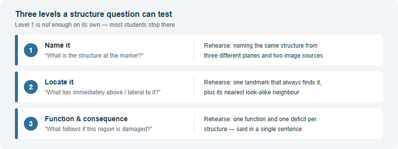
So build every structure card around a landmark relation rather than a description. “Almond-shaped, deep, medial” describes several things. “Sits just in front of the head of the hippocampus, in the medial temporal lobe” finds exactly one. The table below shows the pattern for structures that come up repeatedly, along with the neighbour each is most often confused with — because in practice, a wrong answer is usually the correct structure's neighbour, not a wild guess.
| Structure | The landmark that finds it | Most often confused with |
|---|---|---|
| Thalamus | Paired grey masses either side of the third ventricle, deep and central | Hypothalamus (below it) and the basal ganglia (further lateral) |
| Hippocampus | Curved structure in the medial temporal lobe, along the floor of the temporal horn of the lateral ventricle | Amygdala, which sits immediately anterior to its head |
| Corpus callosum | The large arched white-matter band crossing the midline, obvious on a midsagittal view | Fornix — thinner, and running below the callosum |
| Caudate nucleus | Arcs along the lateral wall of the lateral ventricle, following its curve | Putamen, separated from it by the internal capsule |
| Substantia nigra | Darkly pigmented band within the midbrain, seen on a transverse midbrain section | Cerebral peduncle in front of it; red nucleus nearby |
| Pons | The broad bulge on the ventral brainstem, between midbrain above and medulla below | Medulla, on a cropped or low-contrast image |
| Cerebellum | Tightly foliated cortex below the occipital lobe, behind the brainstem | Occipital cortex, if the foliation is not visible |
Two things to notice about that table. First, every “landmark” column entry is a spatial relation, not an appearance — that is deliberate, because appearance changes with staining, specimen and photograph, while relations do not. Second, the confusion column is where your marks actually live. Drill the pairs, not the singles: hippocampus against amygdala, caudate against putamen, pons against medulla. A structure you can only identify when it appears alone is not yet learned.
A no-lab training loop
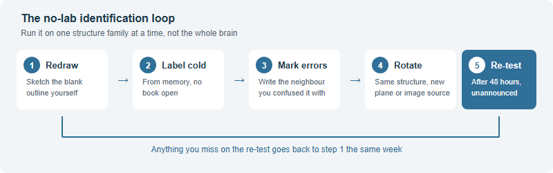
Almost no international-school student in China has routine access to human brain specimens, and you do not need them to make real progress. What you need is a repeatable loop that forces production rather than recognition. Run this on one structure family at a time — brainstem one week, limbic structures the next — rather than on the whole brain at once.
Three details make the difference between running this loop and running it well.
- You must draw the outline yourself. Printing a blank diagram someone else made and filling it in is a recognition task. Sketching the outline forces you to decide where the ventricles, midline and major fissures go, which is exactly the orientation skill the station tests. The drawing does not need to be good.
- Rotate deliberately, not randomly. Work the same structure across coronal, sagittal and transverse views, and across at least two different image sources, before you call it learned. A structure you can only find on the one diagram you revised from is a structure you will lose under pressure.
- Log the confusion, not the miss. “Got hippocampus wrong” tells you nothing next week. “Called the amygdala the hippocampal head, twice, on coronal views” tells you precisely what to drill.
Histology: what a slide is actually asking you to notice
Neurohistology is the half students neglect, usually because it feels less like “the brain” and more like an unfamiliar field of pink and purple. The mental shift that helps is to stop trying to identify the picture and start identifying the features. A slide question is generally asking you to notice some combination of cell shape, cell arrangement, the layering of the tissue, and what the stain has made visible.
A short, practical list of what to build recognition for:
- Neurons against glia. Learn the general appearance of the major glial types — astrocytes, oligodendrocytes, microglia — alongside neurons, rather than learning neurons first and glia “later.” Later never comes, and glia are a large part of the tissue.
- Grey matter against white matter. Cell-body-rich regions look and stain very differently from myelinated fibre tracts. Being able to say which you are looking at, quickly, narrows most questions immediately.
- Distinctive cell shapes and their homes. Purkinje cells in the cerebellar cortex and pyramidal cells in the cerebral cortex and hippocampus are recognisable by shape and by where they sit in the layering — those two facts together are usually enough.
- What a stain shows. Different stains reveal different things: some mark cell bodies, some fill a small number of complete neurons, some highlight myelin. Knowing what a stain is for stops you looking for a feature the slide was never going to show.
Practical note on materials: work from a small number of good images repeatedly rather than a large unsorted collection once. Ten slides you can identify in two seconds beat a hundred you have scrolled past. We also maintain our own gathered pack of past questions and study images for students we work with, though it is worth saying plainly that worked solutions are not available for every year.
What our own cohort suggests, and what to confirm officially
Across one recent season, students prepared by Hanlin took 53 awards at the regional stage, with 31 students advancing to the national round; at the 2026 China national round one teaching group within that cohort took 7 first prizes — including 2nd and 8th in the Junior division and 11th and 13th in the Standard division — along with 11 second prizes and 10 third prizes. Junior (grades 5–8) and Standard (grades 9–12) are China-region divisions rather than an International Brain Bee structure. Those figures are de-identified, reported by Hanlin, describe one cohort in one season, vary by student, and are not a prediction for any individual. They are also not tied to Chinese school admission, which follows separate policy entirely.
The pattern worth passing on is not the count but where the marks moved. Among students who plateaued, the gap was rarely content knowledge; it was that all their anatomy lived in words. The students who improved fastest were the ones who started producing labelled sketches from memory every week instead of rereading chapters — the same principle behind our wider approach to preparing for this competition. Visual recall responds to production, quickly, and it decays just as quickly without it.
Before you plan around anything here, confirm three things officially. Whether the round you are entering includes a specimen or image station at all, and in what form; the division you belong in and the current eligibility rules; and this season's dates, which change every year. For the China round, the official channel is the National Brain Bee Organizing Committee at chinabrainbee.com — and note that Hong Kong, Taiwan and Macao are separate chapters rather than part of the China chapter. Everything stated above about the four World Championship sections and the three-tier structure is documented on thebrainbee.org.
Frequently asked questions
What does the Brain Bee neuroanatomy section involve?
It is one of four World Championship sections, assessing neuroanatomy and neurohistology on real brains or on images rather than by written recall alone.
Can I prepare without access to real specimens?
Yes. Sketch blank outlines yourself, label them cold from memory, then repeat the same structure across different planes and image sources.
Which syllabus material covers the anatomy?
The stated material is Brain Facts, published by the Society for Neuroscience and available at brainfacts.org, or Neuroscience: The Science of the Brain.
Does the China round include a specimen station?
Round formats vary by tier and by year. Confirm with the official China chapter, the National Brain Bee Organizing Committee at chinabrainbee.com.
This is an independent English-language Brain Bee guide operated by Hanlin Education for China-based international-school students. It is not affiliated with, endorsed by, or sponsored by the International Brain Bee (IBB), and it is not the official Brain Bee China national round, which is run by the National Brain Bee Organizing Committee at chinabrainbee.com. Round formats, rules, divisions, eligibility and dates are set by the organisers and change from year to year — always confirm current details on thebrainbee.org before planning around them. Study frameworks, tables and drills described above are our own teaching devices, not published competition rules or marking schemes. Competition results reported here are de-identified, provided by Hanlin, vary by student, are not a prediction of any outcome, and are not tied to Chinese school admission. Confirmed errors are corrected within 7 working days.